 Publications
Publications
2025
Computational design of a high-precision mitochondrial DNA cytosine base editor
Li Mi, Yu-Xuan Li, Xinchen Lv, Zi-Li Wan, Xu Liu, Kairan Zhang, Huican Li, Yue Yao, Leping Zhang, Zhe Xu, Xingyu Zhuang, Kunqian Ji, Min Jiang, Yangming Wang, Peilong Lu
Nature Structural & Molecular Biology, 1-12
Bystander editing remains a major limitation of current base editors, hindering their precision and therapeutic potential. Here, we present a de novo protein design strategy that creates a structurally rigid interface between a DNA-binding TALE domain and a cytosine deaminase, forming a unified editing module termed TALE-oriented deaminase (TOD). Cryo-EM analysis of TOD–DNA complexes confirms that this precise spatial architecture tightly restricts the deaminase activity window, thereby minimizing unwanted deamination. To further enhance editing specificity, we develop a split version, termed DdCBE–TOD, which virtually eliminates off-target editing. As a proof of concept, we apply DdCBE–TOD to generate a mitochondrial disease mouse model and to correct a pathogenic mutation associated with MERRF syndrome in patient-derived cells, achieving single-nucleotide precision. This work introduces a generalizable and computationally guided approach for ultra-precise base editing, offering a promising platform for both mechanistic studies and therapeutic correction of single-nucleotide mutations.
De novo designed voltage-gated anion channels suppress neuron firing
Chen Zhou, Huican Li, Jiaxing Wang, Cheng Qian, Hui Xiong, Zhilin Chu, Qiming Shao, Xuan Li, Shijin Sun, Ke Sun, Aiqin Zhu, Jiawei Wang, Xueqin Jin, Fan Yang, Tamer M Gamal El-Din, Bo Li, Jing Huang, Kun Wu, Peilong Lu
Cell 188 (26), 7495-7511. e21
Design of ion channels responsive to environmental cues has significant implications in modulating cellular activities and sensor development, but it remains a significant challenge due to the complexities involved in designing stimuli-induced conformational changes in proteins. Here, we report the accurate de novo design of voltage-gated anion channels, namely dVGACs. dVGACs adopt a 15-helix pentameric architecture featuring arginine constrictions within the transmembrane span and show voltage-dependent anions currents in patch-clamp experiments. Cryo-electron microscopy (cryo-EM) structures of dVGACs closely align with the design models. Cryo-EM structures and molecular dynamics simulations suggest that the arginine constrictions undergo voltage-induced conformational changes, serving as both a voltage sensor and a selectivity filter as designed. Notably, the anion selectivity and voltage sensitivity of dVGACs can be tuned through targeted mutations for suppressing neuronal firing in situ. The ability to create ion channels with custom-designed conformational changes refreshes our insights into membrane biophysics and unveils diverse potential applications.
De novo design of transmembrane fluorescence-activating proteins
Jingyi Zhu, Mingfu Liang, Ke Sun, Yu Wei, Ruiying Guo, Lijing Zhang, Junhui Shi, Dan Ma, Qi Hu, Gaoxingyu Huang, Peilong Lu
Nature, 1-9
The recognition of ligands by transmembrane proteins is essential for the exchange of materials, energy and information across biological membranes. Progress has been made in the de novo design of transmembrane proteins1,2,3,4,5,6, as well as in designing water-soluble proteins to bind small molecules7,8,9,10,11,12, but de novo design of transmembrane proteins that tightly and specifically bind to small molecules remains an outstanding challenge13. Here we present the accurate design of ligand-binding transmembrane proteins by integrating deep learning and energy-based methods. We designed pre-organized ligand-binding pockets in high-quality four-helix backbones for a fluorogenic ligand, and generated a transmembrane span using gradient-guided hallucination. The designer transmembrane proteins specifically activated fluorescence of the target fluorophore with mid-nanomolar affinity, exhibiting higher brightness and quantum yield compared to those of enhanced green fluorescent protein. These proteins were highly active in the membrane fraction of live bacterial and eukaryotic cells following expression. The crystal and cryogenic electron microscopy structures of the designer protein–ligand complexes were very close to the structures of the design models. We showed that the interactions between ligands and transmembrane proteins within the membrane can be accurately designed. Our work paves the way for the creation of new functional transmembrane proteins, with a wide range of applications including imaging, ligand sensing and membrane transport.
Time-resolved fluorescent proteins expand fluorescent microscopy in temporal and spectral domains
Zizhu Tan, Chia-Heng Hsiung, Jiahui Feng, Yangye Zhang, Yihan Wan, Junlin Chen, Ke Sun, Peilong Lu, Jianyang Zang, Wenxing Yang, Ya Gao, Jiabin Yin, Tong Zhu, Yang Lu, Zijian Pan, Yilong Zou, Can Liao, Xiaosong Li, Yuxuan Ye, Yu Liu, Xin Zhang
Cell 188 (24), 6987-7005. e28
Fluorescence microscopy has been widely applied in the life sciences. While intensity as a steady-state signal is widely used, the time-resolved (tr) signal using fluorescence lifetime remains underexplored. Herein, we present a family of time-resolved fluorescent proteins (tr-FPs) with rationally controlled lifetimes. Using a strategy that regulates lifetime without affecting the spectra of FPs, we have developed a series of tr-FPs that cover the visible spectrum and a wide range of lifetimes. The tr-FPs are employed in temporal-spectral resolved microscopy, allowing for the simultaneous imaging of 9 different proteins in live cells and the correlation of multiple activities to cell cycles. Furthermore, tr-FPs enable multiplexing super-resolution microscopy that concurrently visualizes 4 proteins using the lifetime signal and are demonstrated to quantify the stoichiometry of cellular proteins. Our work introduces the concept and development of tr-FPs as a transformative toolset, presenting opportunities to integrate system complexity and quantitative accuracy into biological research.
Structures and mechanism of the human mitochondrial pyruvate carrier
Jiaming Liang, Junhui Shi, Ailong Song, Meihua Lu, Kairan Zhang, Meng Xu, Gaoxingyu Huang, Peilong Lu, Xudong Wu, Dan Ma
Nature 641 (8061), 258-265
The mitochondrial pyruvate carrier (MPC) is a mitochondrial inner membrane protein complex that is essential for the uptake of pyruvate into the mitochondrial matrix as the primary carbon source for the tricarboxylic acid cycle1,2. Here we present six cryo-electron microscopy structures of human MPC in three states: three structures in the intermembrane space (IMS)-open state, obtained in different conditions; a structure of pyruvate-treated MPC in the occluded state; and two structures in the matrix-facing state, bound with the inhibitor UK5099 or with an inhibitory nanobody on the matrix side. MPC is a heterodimer consisting of MPC1 and MPC2, with the transmembrane domain adopting pseudo-C2 symmetry. Approximate rigid-body movements occur between the IMS-open state and the occluded state, whereas structural changes, mainly on the matrix side, facilitate the transition between the occluded state and the matrix-facing state, revealing an alternating access mechanism during pyruvate transport. In the UK5099-bound structure, the inhibitor fits well and interacts extensively with a pocket that opens to the matrix side. Our findings provide key insights into the mechanisms that underlie MPC-mediated substrate transport, and shed light on the recognition and inhibition of MPC by UK5099, which will facilitate the future development of drugs that target MPC.
Genetically encoding ε-N-methacryllysine into proteins in live cells
Tian-Yi Zhu, Shi-Yi Chen, Mengdi Zhang, Heyu Li, Ting Wu, Emmanuel Ajiboye, Jia Wen Wang, Bi-Kun Jin, Dan-Dan Liu, Xintong Zhou, He Huang, Xiaobo Wan, Ke Sun, Peilong Lu, Yaxin Fu, Ying Yuan, Hai Song, Anna A Sablina, Chao Tong, Long Zhang, Ming Wu, Haifan Wu, Bing Yang
Nature Communications 16 (1), 1-13
Lysine acylation is a ubiquitous post-translational modification (PTM) that plays pivotal roles in various cellular processes, such as transcription, metabolism, protein localization and folding. Thousands of lysine acylation sites have been identified based on advances in antibody enrichment strategies, highly sensitive analysis by mass spectrometry (MS), and bioinformatics. However, only 27 lysine methacrylation (Kmea) sites have been identified exclusively in histone proteins. It is hard to separate, purify and differentiate the Kmea modification from its structural isomer lysine crotonylation (Kcr) using general biochemical approaches. Here, we identify Kmea sites on a non-histone protein, Cyclophillin A (CypA). To investigate the functions of Kmea in CypA, we develop a general genetic code expansion approach to incorporate a non-canonical amino acid (ncAA) ε-N-Methacryllysine (MeaK) into target proteins and identify interacting proteins of methacrylated CypA using affinity-purification MS. We find that Kmea at CypA site 125 regulates cellular redox homeostasis, and HDAC1 is the regulator of Kmea on CypA. Moreover, we discover that genetically encode Kmea can be further methylated to ε-N-methyl-ε-N-methacrylation (Kmemea) in live cells.
Advances in deep learning and generative modeling have transformed the landscape of protein and RNA design, enabling rapid and precise creation of novel biomolecules with tailored structures and functions. In protein design, generative deep learning frameworks now support backbone generation, sequence optimization, and joint sequence–structure co-design with unprecedented accuracy. These approaches have facilitated broad applications ranging from cyclic peptide and non-natural fold engineering to functional tool development, including small-molecule sensing, catalytic center scaffolding, allosteric switching, intracellular logic circuits, and the targeting of intrinsically disordered proteins. Emerging therapeutic applications—such as immune cell engineering, G protein-coupled receptor-targeted miniproteins, receptor-degrading binders, and thermostable antitoxins—demonstrate the translational potential of computational design. Parallel progress in RNA design, driven by enhanced 3D structure prediction models and generative algorithms, is expanding capabilities in aptamer engineering and RNA–protein complexes, despite ongoing challenges in model generalization and experimental validation. Together, these developments highlight a new era of AI-driven molecular engineering, in which unified protein–RNA modeling, large-scale sampling, and automated experimental pipelines will accelerate the creation of programmable biological systems and next-generation therapeutics.
Harnessing advances in artificial intelligence for protein design
Russell Johnson
Nat Chem Biol 22, 1–4 (2026)
Machine learning-based tools have revolutionized how scientists study protein structure. Here, Nature Chemical Biology speaks to Cecilia Clementi, Bruno Correia and Peilong Lu about progress in developing computational tools for predicting protein structure and properties, how these programs can be used for protein design, and the developments they would like to see in the field.
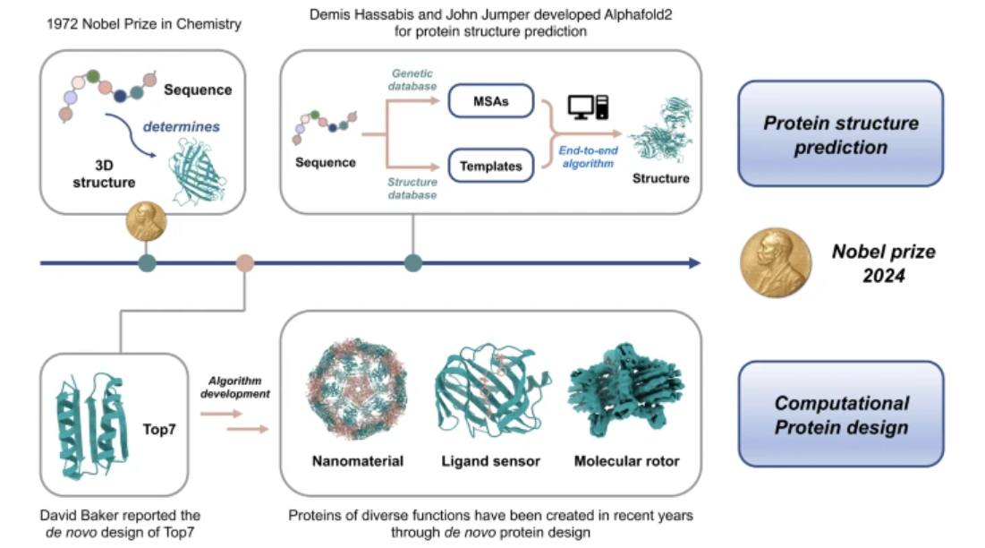
Computational protein design and structure prediction—The 2024 Nobel Prize in Chemistry
Peilong Lu, Lei Liu
Science China Chemistry 68 (3), 812-814
2024
Accurate de novo design of heterochiral protein–protein interactions
Ke Sun, Sicong Li, Bowen Zheng, Yanlei Zhu, Tongyue Wang, Mingfu Liang, Yue Yao, Kairan Zhang, Jizhong Zhang, Hongyong Li, Dongyang Han, Jishen Zheng, Brian Coventry, Longxing Cao, David Baker, Lei Liu, Peilong Lu
Cell Research 34 (12), 846-858
Abiotic d-proteins that selectively bind to natural l-proteins have gained significant biotechnological interest. However, the underlying structural principles governing such heterochiral protein–protein interactions remain largely unknown. In this study, we present the de novo design of d-proteins consisting of 50–65 residues, aiming to target specific surface regions of l-proteins or l-peptides. Our designer d-protein binders exhibit nanomolar affinity toward an artificial l-peptide, as well as two naturally occurring proteins of therapeutic significance: the D5 domain of human tropomyosin receptor kinase A (TrkA) and human interleukin-6 (IL-6). Notably, these d-protein binders demonstrate high enantiomeric specificity and target specificity. In cell-based experiments, designer d-protein binders effectively inhibited the downstream signaling of TrkA and IL-6 with high potency. Moreover, these binders exhibited remarkable thermal stability and resistance to protease degradation. Crystal structure of the designed heterochiral d-protein–l-peptide complex, obtained at a resolution of 2.0 Å, closely resembled the design model, indicating that the computational method employed is highly accurate. Furthermore, the crystal structure provides valuable information regarding the interactions between helical l-peptides and d-proteins, particularly elucidating a novel mode of heterochiral helix–helix interactions. Leveraging the design of d-proteins specifically targeting l-peptides or l-proteins opens up avenues for systematic exploration of the mirror-image protein universe, paving the way for a diverse range of applications.
Xinchen Lv, Yuanyuan Zhang, Ke Sun, Qi Yang, Jianhua Luo, Liang Tao, Peilong Lu
Nature Communications 15 (1), 8521
Clostridioides difficile toxin B (TcdB) is the key virulence factor accounting for C. difficile infection-associated symptoms. Effectively neutralizing different TcdB variants with a universal solution poses a significant challenge. Here we present the de novo design and characterization of pan-specific mini-protein binders against major TcdB subtypes. Our design successfully binds to the first receptor binding interface (RBI-1) of the varied TcdB subtypes, exhibiting affinities ranging from 20 pM to 10 nM. The cryo-electron microscopy (cryo-EM) structures of the mini protein binder in complex with TcdB1 and TcdB4 are consistent with the computational design models. The engineered and evolved variants of the mini-protein binder and chondroitin sulfate proteoglycan 4 (CSPG4), another natural receptor that binds to the second RBI (RBI-2) of TcdB, better neutralize major TcdB variants both in cells and in vivo, as demonstrated by the colon-loop assay using female mice. Our findings provide valuable starting points for the development of therapeutics targeting C. difficile infections (CDI).
Justin A Peruzzi, Taylor F Gunnels, Hailey I Edelstein, Peilong Lu, David Baker, Joshua N Leonard, Neha P Kamat
Nature communications 15 (1), 5618
Naturally generated lipid nanoparticles termed extracellular vesicles (EVs) hold significant promise as engineerable therapeutic delivery vehicles. However, active loading of protein cargo into EVs in a manner that is useful for delivery remains a challenge. Here, we demonstrate that by rationally designing proteins to traffic to the plasma membrane and associate with lipid rafts, we can enhance loading of protein cargo into EVs for a set of structurally diverse transmembrane and peripheral membrane proteins. We then demonstrate the capacity of select lipid tags to mediate increased EV loading and functional delivery of an engineered transcription factor to modulate gene expression in target cells. We envision that this technology could be leveraged to develop new EV-based therapeutics that deliver a wide array of macromolecular cargo.
Dan-Dan Liu, Wenlong Ding, Jin-Tao Cheng, Qiushi Wei, Yinuo Lin, Tian-Yi Zhu, Jing Tian, Ke Sun, Long Zhang, Peilong Lu, Fan Yang, Chao Liu, Shibing Tang, Bing Yang
Nature Communications 15 (1), 5221
Latent bioreactive unnatural amino acids (Uaas) have been widely used in the development of covalent drugs and identification of protein interactors, such as proteins, DNA, RNA and carbohydrates. However, it is challenging to perform high-throughput identification of Uaa cross-linking products due to the complexities of protein samples and the data analysis processes. Enrichable Uaas can effectively reduce the complexities of protein samples and simplify data analysis, but few cross-linked peptides were identified from mammalian cell samples with these Uaas. Here we develop an enrichable and multiple amino acids reactive Uaa, eFSY, and demonstrate that eFSY is MS cleavable when eFSY-Lys and eFSY-His are the cross-linking products. An identification software, AixUaa is developed to decipher eFSY mass cleavable data. We systematically identify direct interactomes of Thioredoxin 1 (Trx1) and Selenoprotein M (SELM) with eFSY and AixUaa.
Hydrophobic mismatch drives self-organization of designer proteins into synthetic membranes
Justin A Peruzzi, Jan Steinkühler, Timothy Q Vu, Taylor F Gunnels, Vivian T Hu, Peilong Lu, David Baker, Neha P Kamat
Nature communications 15 (1), 3162
The organization of membrane proteins between and within membrane-bound compartments is critical to cellular function. Yet we lack approaches to regulate this organization in a range of membrane-based materials, such as engineered cells, exosomes, and liposomes. Uncovering and leveraging biophysical drivers of membrane protein organization to design membrane systems could greatly enhance the functionality of these materials. Towards this goal, we use de novo protein design, molecular dynamic simulations, and cell-free systems to explore how membrane-protein hydrophobic mismatch could be used to tune protein cotranslational integration and organization in synthetic lipid membranes. We find that membranes must deform to accommodate membrane-protein hydrophobic mismatch, which reduces the expression and co-translational insertion of membrane proteins into synthetic membranes. We use this principle to sort proteins both between and within membranes, thereby achieving one-pot assembly of vesicles with distinct functions and controlled split-protein assembly, respectively. Our results shed light on protein organization in biological membranes and provide a framework to design self-organizing membrane-based materials with applications such as artificial cells, biosensors, and therapeutic nanoparticles.
AI accurately predicting the structure of biomolecular interactions
Zhenling Peng, Peilong Lu, Jianyi Yang
Cell Research 34 (9), 601-602
In two recent studies published in Nature and Science, researchers successfully developed unified AI architectures capable of predicting the structures of all biomolecules. This remarkable advancement is expected to have a profound impact on future biomedical research and drug design by offering crucial information regarding the interactions that govern both physiological and pathological processes.
2023
Birte Höcker, Peilong Lu, Anum Glasgow, Debora S Marks, Pranam Chatterjee, Joanna SG Slusky, Ora Schueler-Furman, Possu Huang
Cell Systems 14 (8), 629-632
Easy access and critical use It is important to make novel technologies easily accessible to non-expert users and provide guidance for their use. The protein design community openly shares methods, programs, and source code and has done so even before the AI (r) evolution. What changed is the scale and increased interest due to the improved precision in predictions. In fact, protein structures have already been predicted at large scale, and biologists can simply access them in the AlphaFold Protein Structure Database or ESM Metagenomic Atlas. Due to these incredibly useful resources, they do not even have to run the predictions themselves.
For protein design applications, though, the issue differs. Aims vary and approaches need to be adjusted based on the individual design goal. Again, many open-source tools are available that are constantly updated and diversified. Open-science platforms such as …
2022
De novo design of membrane transport proteins
Chen Zhou, Peilong Lu
Proteins: Structure, Function, and Bioinformatics 90 (10), 1800-1806
Membrane transport proteins, which include transporters and channels, are delicate protein machineries that mediate the exchange of a variety of substances across biomembranes. Accumulated structural and functional knowledge allows for the de novo design of transport proteins with new structures that do not exist in nature. Analysis based on these novel proteins provides new insights into the principles that govern protein assembly, conformational change, and substrate recognition. Here, we review the advances in the de novo design of transporters and channels over recent years and highlight the challenges and opportunities in this field.
Computational design of transmembrane proteins
Jingyi Zhu, Peilong Lu
Current opinion in structural biology 74, 102381
In recent decades, major progress has been made in the design of water-soluble proteins, yet the design of transmembrane proteins has lagged considerably. Despite their biological and pharmaceutical importance, only a limited number of transmembrane proteins have been successfully designed owing to the complexity of the membrane environment and difficulties in experimental characterization. Here, we introduce principles for transmembrane protein design in general and discuss design examples, including scaffold proteins and functional proteins. We also discuss how developments in design methods have advanced the field and what we may achieve with recent breakthroughs in structural biology.
Justin Peruzzi, Jan Steinkuehler, Timothy Vu, Peilong Lu, David Baker, Neha Kamat
Biophysical Journal 121 (3), 313a
Cell-mimetic membranes are powerful tools for studying lipid-protein interactions and engineering membrane-based materials. Many proteins have been incorporated into artificial membranes, however the ability to organize proteins between and within synthetic membranes has not yet been achieved. Cells possess many membrane-bound structures, each unique in lipid and protein composition, enabling diverse and specialized functions. Furthermore, biological membranes are laterally heterogenous, possessing lipid rafts which enable distinct regions differing in composition, biophysical properties, and function. Controlling where membrane proteins localize in artificial membranes is a first step to creating more accurate cell-mimetic membranes capable of performing complex biochemical processes. In cells, lipid-protein interactions drive inter- and intra-membrane protein organization. Inspired by this, we investigated how hydrophobic mismatch between synthetic proteins and membranes could be leveraged to spatially organize proteins between and within synthetic membranes. First, we characterized how hydrophobic mismatch affects the insertion and folding of cell-free expressed membrane proteins into synthetic membranes. We next investigated how membrane-protein hydrophobic mismatch may be used as a handle to control inter- and intra-membrane organization through experimental and computational analysis. We demonstrate that this organization enables the assembly of vesicles with distinct functions, leading to the controlled release of encapsulated cargo and the segregation of membrane-bound enzymes within distinct lipid domains. This study highlights how lipid-protein interactions may be leveraged in cell-free systems to assemble more complex cell-mimetic structures and demonstrates how the organization of proteins within synthetic membranes may enable more complex behaviors to be studied within artificial membranes.
2021
Cryo-EM structure of human Wntless in complex with Wnt3a
Qing Zhong, Yanyu Zhao, Fangfei Ye, Zaiyu Xiao, Gaoxingyu Huang, Meng Xu, Yuanyuan Zhang, Xiechao Zhan, Ke Sun, Zhizhi Wang, Shanshan Cheng, Shan Feng, Xiuxiu Zhao, Jizhong Zhang, Peilong Lu, Wenqing Xu, Qiang Zhou, Dan Ma
Nature communications 12 (1), 4541
Wntless (WLS), an evolutionarily conserved multi-pass transmembrane protein, is essential for secretion of Wnt proteins. Wnt-triggered signaling pathways control many crucial life events, whereas aberrant Wnt signaling is tightly associated with many human diseases including cancers. Here, we report the cryo-EM structure of human WLS in complex with Wnt3a, the most widely studied Wnt, at 2.2 Å resolution. The transmembrane domain of WLS bears a GPCR fold, with a conserved core cavity and a lateral opening. Wnt3a interacts with WLS at multiple interfaces, with the lipid moiety on Wnt3a traversing a hydrophobic tunnel of WLS transmembrane domain and inserting into membrane. A β-hairpin of Wnt3a containing the conserved palmitoleoylation site interacts with WLS extensively, which is crucial for WLS-mediated Wnt secretion. The flexibility of the Wnt3a loop/hairpin regions involved in the multiple binding sites indicates induced fit might happen when Wnts are bound to different binding partners. Our findings provide important insights into the molecular mechanism of Wnt palmitoleoylation, secretion and signaling.
Parisa Hosseinzadeh, Paris R Watson, Timothy W Craven, Xinting Li, Stephen Rettie, Fátima Pardo-Avila, Asim K Bera, Vikram Khipple Mulligan, Peilong Lu, Alexander S Ford, Brian D Weitzner, Lance J Stewart, Adam P Moyer, Maddalena Di Piazza, Joshua G Whalen, Per Jr Greisen, David W Christianson, David Baker
Nature Communications 12 (1), 3384
Despite recent success in computational design of structured cyclic peptides, de novo design of cyclic peptides that bind to any protein functional site remains difficult. To address this challenge, we develop a computational “anchor extension” methodology for targeting protein interfaces by extending a peptide chain around a non-canonical amino acid residue anchor. To test our approach using a well characterized model system, we design cyclic peptides that inhibit histone deacetylases 2 and 6 (HDAC2 and HDAC6) with enhanced potency compared to the original anchor (IC50 values of 9.1 and 4.4 nM for the best binders compared to 5.4 and 0.6 µM for the anchor, respectively). The HDAC6 inhibitor is among the most potent reported so far. These results highlight the potential for de novo design of high-affinity protein-peptide interfaces, as well as the challenges that remain.
2020
Computational design of transmembrane pores
Chunfu Xu, Peilong Lu, Tamer M Gamal El-Din, Xue Y Pei, Matthew C Johnson, Atsuko Uyeda, Matthew J Bick, Qi Xu, Daohua Jiang, Hua Bai, Gabriella Reggiano, Yang Hsia, TJ Brunette, Jiayi Dou, Dan Ma, Eric M Lynch, Scott E Boyken, Po-Ssu Huang, Lance Stewart, Frank DiMaio, Justin M Kollman, Ben F Luisi, Tomoaki Matsuura, William A Catterall, David Baker
Nature 585 (7823), 129-134
Transmembrane channels and pores have key roles in fundamental biological processes1 and in biotechnological applications such as DNA nanopore sequencing2,3,4, resulting in considerable interest in the design of pore-containing proteins. Synthetic amphiphilic peptides have been found to form ion channels5,6, and there have been recent advances in de novo membrane protein design7,8 and in redesigning naturally occurring channel-containing proteins9,10. However, the de novo design of stable, well-defined transmembrane protein pores that are capable of conducting ions selectively or are large enough to enable the passage of small-molecule fluorophores remains an outstanding challenge11,12. Here we report the computational design of protein pores formed by two concentric rings of α-helices that are stable and monodisperse in both their water-soluble and their transmembrane forms. Crystal structures of the water-soluble forms of a 12-helical pore and a 16-helical pore closely match the computational design models. Patch-clamp electrophysiology experiments show that, when expressed in insect cells, the transmembrane form of the 12-helix pore enables the passage of ions across the membrane with high selectivity for potassium over sodium; ion passage is blocked by specific chemical modification at the pore entrance. When incorporated into liposomes using in vitro protein synthesis, the transmembrane form of the 16-helix pore—but not the 12-helix pore—enables the passage of biotinylated Alexa Fluor 488. A cryo-electron microscopy structure of the 16-helix transmembrane pore closely matches the design model. The ability to produce structurally and functionally well-defined transmembrane pores opens the door to the creation of designer channels and pores for a wide variety of applications.
When de novo-designed protein logics meet CAR-T therapies
Mingqi Xie, Peilong Lu
Nature 585 (7823), 129-134
Protein logic gates acting at posttranscriptional levels are unique in being amenable to the control of both intracellular and intercellular biological processes. In a recent paper published in Science, Lajoie et al. engineered highly versatile colocalization-dependent protein switch that are activated upon detection of logic combinations (eg, AND, OR and NOT) of specific cell surface antigens, providing another important asset for “universal” CAR-T therapies.
In recent years, chimeric antigen receptor T cell (CAR-T) therapies based on adoptive transfer of ex vivo engineered patient-specific T cells have finally arrived on the central stage of the pharmaceutical industry. 1 Despite showing excellent response rates in many patients suffering from blood tumors (eg, leukemia), many hurdles still need to be overcome to accelerate the development of future CAR-T therapies. 2 One major challenge is that, in many cases, multiple …
Publications Prior to Establishing the Lab
2019
Programmable design of orthogonal protein heterodimers
Zibo Chen, Scott E Boyken, Mengxuan Jia, Florian Busch, David Flores-Solis, Matthew J Bick, Peilong Lu, Zachary L VanAernum, Aniruddha Sahasrabuddhe, Robert A Langan, Sherry Bermeo, TJ Brunette, Vikram Khipple Mulligan, Lauren P Carter, Frank DiMaio, Nikolaos G Sgourakis, Vicki H Wysocki, David Baker
Nature 565 (7737), 106-111
Specificity of interactions between two DNA strands, or between protein and DNA, is often achieved by varying bases or side chains coming off the DNA or protein backbone—for example, the bases participating in Watson–Crick pairing in the double helix, or the side chains contacting DNA in TALEN–DNA complexes. By contrast, specificity of protein–protein interactions usually involves backbone shape complementarity1, which is less modular and hence harder to generalize. Coiled-coil heterodimers are an exception, but the restricted geometry of interactions across the heterodimer interface (primarily at the heptad a and d positions2) limits the number of orthogonal pairs that can be created simply by varying side-chain interactions3,4. Here we show that protein–protein interaction specificity can be achieved using extensive and modular side-chain hydrogen-bond networks. We used the Crick generating equations5 to produce millions of four-helix backbones with varying degrees of supercoiling around a central axis, identified those accommodating extensive hydrogen-bond networks, and used Rosetta to connect pairs of helices with short loops and to optimize the remainder of the sequence. Of 97 such designs expressed in Escherichia coli, 65 formed constitutive heterodimers, and the crystal structures of four designs were in close agreement with the computational models and confirmed the designed hydrogen-bond networks. In cells, six heterodimers were fully orthogonal, and in vitro—following mixing of 32 chains from 16 heterodimer designs, denaturation in 5 M guanidine hydrochloride and reannealing—almost all of the interactions observed by native mass spectrometry were between the designed cognate pairs. The ability to design orthogonal protein heterodimers should enable sophisticated protein-based control logic for synthetic biology, and illustrates that nature has not fully explored the possibilities for programmable biomolecular interaction modalities.
2018
Crystal structure of a membrane-bound O-acyltransferase
Dan Ma, Zhizhi Wang, Christopher N Merrikh, Kevin S Lang, Peilong Lu, Xin Li, Houra Merrikh, Zihe Rao, Wenqing Xu
Nature 562 (7726), 286-290
Membrane-bound O-acyltransferases (MBOATs) are a superfamily of integral transmembrane enzymes that are found in all kingdoms of life1. In bacteria, MBOATs modify protective cell-surface polymers. In vertebrates, some MBOAT enzymes—such as acyl-coenzyme A:cholesterol acyltransferase and diacylglycerol acyltransferase 1—are responsible for lipid biosynthesis or phospholipid remodelling2,3. Other MBOATs, including porcupine, hedgehog acyltransferase and ghrelin acyltransferase, catalyse essential lipid modifications of secreted proteins such as Wnt, hedgehog and ghrelin, respectively4,5,6,7,8,9,10. Although many MBOAT proteins are important drug targets, little is known about their molecular architecture and functional mechanisms. Here we present crystal structures of DltB, an MBOAT responsible for the d-alanylation of cell-wall teichoic acid in Gram-positive bacteria11,12,13,14,15,16, both alone and in complex with the d-alanyl donor protein DltC. DltB contains a ring of 11 peripheral transmembrane helices, which shield a highly conserved extracellular structural funnel extending into the middle of the lipid bilayer. The conserved catalytic histidine residue is located at the bottom of this funnel and is connected to the intracellular DltC through a narrow tunnel. Mutation of either the catalytic histidine or the DltC-binding site of DltB abolishes the d-alanylation of lipoteichoic acid and sensitizes the Gram-positive bacterium Bacillus subtilis to cell-wall stress, which suggests cross-membrane catalysis involving the tunnel. Structure-guided sequence comparison among DltB and vertebrate MBOATs reveals a conserved structural core and suggests that MBOATs from different organisms have similar catalytic mechanisms. Our structures provide a template for understanding structure–function relationships in MBOATs and for developing therapeutic MBOAT inhibitors.
Structural basis for gating pore current in periodic paralysis
Daohua Jiang, Tamer M Gamal El-Din, Christopher Ing, Peilong Lu, Regis Pomes, Ning Zheng, William A Catterall
Nature 557 (7706), 590-594
Potassium-sensitive hypokalaemic and normokalaemic periodic paralysis are inherited skeletal muscle diseases characterized by episodes of flaccid muscle weakness1,2. They are caused by single mutations in positively charged residues (‘gating charges’) in the S4 transmembrane segment of the voltage sensor of the voltage-gated sodium channel Nav1.4 or the calcium channel Cav1.11,2. Mutations of the outermost gating charges (R1 and R2) cause hypokalaemic periodic paralysis1,2 by creating a pathogenic gating pore in the voltage sensor through which cations leak in the resting state3,4. Mutations of the third gating charge (R3) cause normokalaemic periodic paralysis5 owing to cation leak in both activated and inactivated states6. Here we present high-resolution structures of the model bacterial sodium channel NavAb with the analogous gating-charge mutations7,8, which have similar functional effects as in the human channels. The R2G and R3G mutations have no effect on the backbone structures of the voltage sensor, but they create an aqueous cavity near the hydrophobic constriction site that controls gating charge movement through the voltage sensor. The R3G mutation extends the extracellular aqueous cleft through the entire length of the activated voltage sensor, creating an aqueous path through the membrane. Conversely, molecular modelling shows that the R2G mutation creates a continuous aqueous path through the membrane only in the resting state. Crystal structures of NavAb(R2G) in complex with guanidinium define a potential drug target site. Molecular dynamics simulations illustrate the mechanism of Na+ permeation through the mutant gating pore in concert with conformational fluctuations of the gating charge R4. Our results reveal pathogenic mechanisms of periodic paralysis at the atomic level and suggest designs of drugs that may prevent ionic leak and provide symptomatic relief from hypokalaemic and normokalaemic periodic paralysis.
Accurate computational design of multipass transmembrane proteins
Peilong Lu, Duyoung Min, Frank DiMaio, Kathy Y Wei, Michael D Vahey, Scott E Boyken, Zibo Chen, Jorge A Fallas, George Ueda, William Sheffler, Vikram Khipple Mulligan, Wenqing Xu, James U Bowie, David Baker
Science 359 (6379), 1042-1046
The computational design of transmembrane proteins with more than one membrane-spanning region remains a major challenge. We report the design of transmembrane monomers, homodimers, trimers, and tetramers with 76 to 215 residue subunits containing two to four membrane-spanning regions and up to 860 total residues that adopt the target oligomerization state in detergent solution. The designed proteins localize to the plasma membrane in bacteria and in mammalian cells, and magnetic tweezer unfolding experiments in the membrane indicate that they are very stable. Crystal structures of the designed dimer and tetramer—a rocket-shaped structure with a wide cytoplasmic base that funnels into eight transmembrane helices—are very close to the design models. Our results pave the way for the design of multispan membrane proteins with new functions.
2015
An atomic structure of human γ-secretase
Xiao-chen Bai, Chuangye Yan, Guanghui Yang, Peilong Lu, Dan Ma, Linfeng Sun, Rui Zhou, Sjors HW Scheres, Yigong Shi
Dysfunction of the intramembrane protease γ-secretase is thought to cause Alzheimer’s disease, with most mutations derived from Alzheimer’s disease mapping to the catalytic subunit presenilin 1 (PS1). Here we report an atomic structure of human γ-secretase at 3.4 Å resolution, determined by single-particle cryo-electron microscopy. Mutations derived from Alzheimer’s disease affect residues at two hotspots in PS1, each located at the centre of a distinct four transmembrane segment (TM) bundle. TM2 and, to a lesser extent, TM6 exhibit considerable flexibility, yielding a plastic active site and adaptable surrounding elements. The active site of PS1 is accessible from the convex side of the TM horseshoe, suggesting considerable conformational changes in nicastrin extracellular domain after substrate recruitment. Component protein APH-1 serves as a scaffold, anchoring the lone transmembrane helix from nicastrin and supporting the flexible conformation of PS1. Ordered phospholipids stabilize the complex inside the membrane. Our structure serves as a molecular basis for mechanistic understanding of γ-secretase function.
Nature 525 (7568), 212-217
2014
Crystal structure of the γ-secretase component nicastrin
Tian Xie, Chuangye Yan, Rui Zhou, Yanyu Zhao, Linfeng Sun, Guanghui Yang, Peilong Lu, Dan Ma, Yigong Shi
Proceedings of the National Academy of Sciences 111 (37), 13349-13354
γ-Secretase is an intramembrane protease responsible for the generation of amyloid-β (Aβ) peptides. Aberrant accumulation of Aβ leads to the formation of amyloid plaques in the brain of patients with Alzheimer's disease. Nicastrin is the putative substrate-recruiting component of the γ-secretase complex. No atomic-resolution structure had been identified on γ-secretase or any of its four components, hindering mechanistic understanding of γ-secretase function. Here we report the crystal structure of nicastrin from Dictyostelium purpureum at 1.95-Å resolution. The extracellular domain of nicastrin contains a large lobe and a small lobe. The large lobe of nicastrin, thought to be responsible for substrate recognition, associates with the small lobe through a hydrophobic pivot at the center. The putative substrate-binding pocket is shielded from the small lobe by a lid, which blocks substrate entry. These structural features suggest a working model of nicastrin function. Analysis of nicastrin structure provides insights into the assembly and architecture of the γ-secretase complex.
Three-dimensional structure of human γ-secretase
Peilong Lu, Xiao-chen Bai, Dan Ma, Tian Xie, Chuangye Yan, Linfeng Sun, Guanghui Yang, Yanyu Zhao, Rui Zhou, Sjors HW Scheres, Yigong Shi
Nature 512 (7513), 166-170
The γ-secretase complex, comprising presenilin 1 (PS1), PEN-2, APH-1 and nicastrin, is a membrane-embedded protease that controls a number of important cellular functions through substrate cleavage. Aberrant cleavage of the amyloid precursor protein (APP) results in aggregation of amyloid-β, which accumulates in the brain and consequently causes Alzheimer’s disease. Here we report the three-dimensional structure of an intact human γ-secretase complex at 4.5 Å resolution, determined by cryo-electron-microscopy single-particle analysis. The γ-secretase complex comprises a horseshoe-shaped transmembrane domain, which contains 19 transmembrane segments (TMs), and a large extracellular domain (ECD) from nicastrin, which sits immediately above the hollow space formed by the TM horseshoe. Intriguingly, nicastrin ECD is structurally similar to a large family of peptidases exemplified by the glutamate carboxypeptidase PSMA. This structure serves as an important basis for understanding the functional mechanisms of the γ-secretase complex.
Structure and mechanism of a eukaryotic transmembrane ascorbate-dependent oxidoreductase
Peilong Lu, Dan Ma, Chuangye Yan, Xinqi Gong, Mingjian Du, Yigong Shi
Proceedings of the National Academy of Sciences 111 (5), 1813-1818
Vitamin C, also known as ascorbate, is required in numerous essential metabolic reactions in eukaryotes. The eukaryotic ascorbate-dependent oxidoreductase cytochrome b561 (Cyt b561), a family of highly conserved transmembrane enzymes, plays an important role in ascorbate recycling and iron absorption. Although Cyt b561 was identified four decades ago, its atomic structure and functional mechanism remain largely unknown. Here, we report the high-resolution crystal structures of cytochrome b561 from Arabidopsis thaliana in both substrate-free and substrate-bound states. Cyt b561 forms a homodimer, with each protomer consisting of six transmembrane helices and two heme groups. The negatively charged substrate ascorbate, or monodehydroascorbate, is enclosed in a positively charged pocket on either side of the membrane. Two highly conserved amino acids, Lys81 and His106, play an essential role in substrate recognition and catalysis. Our structural and biochemical analyses allow the proposition of a general electron transfer mechanism for members of the Cyt b561 family.
2013
Dan Ma, Peilong Lu, Yigong Shi
GadC, a central component of the Escherichia coli acid resistance system, is a Glu/GABA antiporter. A previous structural study and biochemical characterization showed that GadC exhibits a stringent pH dependence for substrate transport, with no detectable activity at pH values above 6.5. However, the substrate selectivity and the mechanism of pH-dependent transport activity of GadC remain enigmatic. In this study, we demonstrate that GadC selectively transports Glu with no net charge and GABA with a positive charge. A C-plug-truncated variant of GadC (residues 1–470) transported Gln (a mimic of Glu with no net charge), but not Glu, even at pH 8.0. The pH-dependent transport of Gln by this GadC variant was shifted ∼1 unit toward a higher pH compared with Glu transport. Taken together, the results identify the substrate selectivity for GadC and show that the protonation states of substrates are crucial for transport.
Journal of Biological Chemistry 288 (21), 15148-15153
L-glutamine provides acid resistance for Escherichia coli through enzymatic release of ammonia
Peilong Lu, Dan Ma, Yuling Chen, Yingying Guo, Guo-Qiang Chen, Haiteng Deng, Yigong Shi
Cell research 23 (5), 635-644
Bacteria, exemplified by enteropathogenic Escherichia coli (E. coli), rely on elaborate acid resistance systems to survive acidic environment (such as the stomach). Comprehensive understanding of bacterial acid resistance is important for prevention and clinical treatment. In this study, we report a previously uncharacterized type of acid resistance system in E. coli that relies on L-glutamine (Gln), one of the most abundant food-borne free amino acids. Upon uptake into E. coli, Gln is converted to L-glutamate (Glu) by the acid-activated glutaminase YbaS, with concomitant release of gaseous ammonia. The free ammonia neutralizes proton, resulting in elevated intracellular pH under acidic environment. We show that YbaS and the amino acid antiporter GadC, which exchanges extracellular Gln with intracellular Glu, together constitute an acid resistance system that is sufficient for E. coli survival under extremely acidic environment.
2012
Structure and mechanism of a glutamate–GABA antiporter
Dan Ma, Peilong Lu, Chuangye Yan, Chao Fan, Ping Yin, Jiawei Wang, Yigong Shi
Nature 483 (7391), 632-636
Food-borne hemorrhagic Escherichia coli, exemplified by the strains O157:H7 and O104:H4 (refs 1, 2), require elaborate acid-resistance systems (ARs)3 to survive the extremely acidic environment such as the stomach (pH ≈ 2). AR2 expels intracellular protons through the decarboxylation of l-glutamate (Glu) in the cytoplasm and exchange of the reaction product γ-aminobutyric acid (GABA) with extracellular Glu. The latter process is mediated by the Glu–GABA antiporter GadC4,5, a representative member of the amino-acid–polyamine–organocation superfamily of membrane transporters. The functional mechanism of GadC remains largely unknown. Here we show, with the use of an in vitro proteoliposome-based assay, that GadC transports GABA/Glu only under acidic conditions, with no detectable activity at pH values higher than 6.5. We determined the crystal structure of E. coli GadC at 3.1 Å resolution under basic conditions. GadC, comprising 12 transmembrane segments (TMs), exists in a closed state, with its carboxy-terminal domain serving as a plug to block an otherwise inward-open conformation. Structural and biochemical analyses reveal the essential transport residues, identify the transport path and suggest a conserved transport mechanism involving the rigid-body rotation of a helical bundle for GadC and other amino acid antiporters.
Crystal structure and biochemical analyses reveal Beclin 1 as a novel membrane binding protein
Weijiao Huang, Wooyoung Choi, Wanqiu Hu, Na Mi, Qiang Guo, Meisheng Ma, Mei Liu, Yuan Tian, Peilong Lu, Feng-Liang Wang, Haiteng Deng, Lei Liu, Ning Gao, Li Yu, Yigong Shi
Cell research 22 (3), 473-489
The Beclin 1 gene is a haplo-insufficient tumor suppressor and plays an essential role in autophagy. However, the molecular mechanism by which Beclin 1 functions remains largely unknown. Here we report the crystal structure of the evolutionarily conserved domain (ECD) of Beclin 1 at 1.6 Å resolution. Beclin 1 ECD exhibits a previously unreported fold, with three structural repeats arranged symmetrically around a central axis. Beclin 1 ECD defines a novel class of membrane-binding domain, with a strong preference for lipid membrane enriched with cardiolipin. The tip of a surface loop in Beclin 1 ECD, comprising three aromatic amino acids, acts as a hydrophobic finger to associate with lipid membrane, consequently resulting in the deformation of membrane and liposomes. Mutation of these aromatic residues rendered Beclin 1 unable to stably associate with lipid membrane in vitro and unable to fully rescue autophagy in Beclin 1-knockdown cells in vivo. These observations form an important framework for deciphering the biological functions of Beclin 1.
Structure of the mRNA splicing complex component Cwc2: insights into RNA recognition
Peilong Lu, Guifeng Lu, Chuangye Yan, Li Wang, Wenqi Li, Ping Yin
Biochemical Journal 441 (2), 591-597
The Prp19-associated complex [NTC (nineteen complex)] plays a crucial role in intron removal during premature mRNA splicing in eukaryotes. Only one component of the NTC, Cwc2, is capable of binding RNA. In the present study we report the 1.9 Å (1 Å=0.1 nm) X-ray structure of the Cwc2 core domain, which is both necessary and sufficient for RNA binding. The Cwc2 core domain contains two sub-domains, a CCCH-type ZnF (zinc finger) and a RRM (RNA recognition motif). Unexpectedly, the ZnF domain and the RRM form a single folding unit, glued together by extensive hydrophobic interactions and hydrogen bonds. Structure-guided mutational analysis revealed that the intervening loop [known as the RB loop (RNA-binding loop)] between ZnF and RRM plays an essential role in RNA binding. In addition, a number of highly conserved positively charged residues on the β-strands of RRM make an important contribution to RNA binding. Intriguingly, these residues and a portion of the RB loop constitute an extended basic surface strip that encircles Cwc2 halfway. The present study serves as a framework for understanding the regulatory function of the NTC in RNA splicing.
2009
Structure of the formate transporter FocA reveals a pentameric aquaporin-like channel
Yi Wang, Yongjian Huang, Jiawei Wang, Chao Cheng, Weijiao Huang, Peilong Lu, Ya-Nan Xu, Pengye Wang, Nieng Yan, Yigong Shi
Nature 462 (7272), 467-472
FocA is a representative member of the formate–nitrite transporter family, which transports short-chain acids in bacteria, archaea, fungi, algae and parasites. The structure and transport mechanism of the formate–nitrite transporter family remain unknown. Here we report the crystal structure of Escherichia coli FocA at 2.25 Å resolution. FocA forms a symmetric pentamer, with each protomer consisting of six transmembrane segments. Despite a lack of sequence homology, the overall structure of the FocA protomer closely resembles that of aquaporin and strongly argues that FocA is a channel, rather than a transporter. Structural analysis identifies potentially important channel residues, defines the channel path and reveals two constriction sites. Unlike aquaporin, FocA is impermeable to water but allows the passage of formate. A structural and biochemical investigation provides mechanistic insights into the channel activity of FocA.